
HUMAN DIGESTIVE ANATOMY
Understanding the anatomy of the stomach
The stomach is a muscular, J-shaped part of the digestive tract located between the oesophagus and the duodenum. It acts as a temporary reservoir for food, mixes food with gastric secretions and gradually releases the resulting mixture into the small intestine.
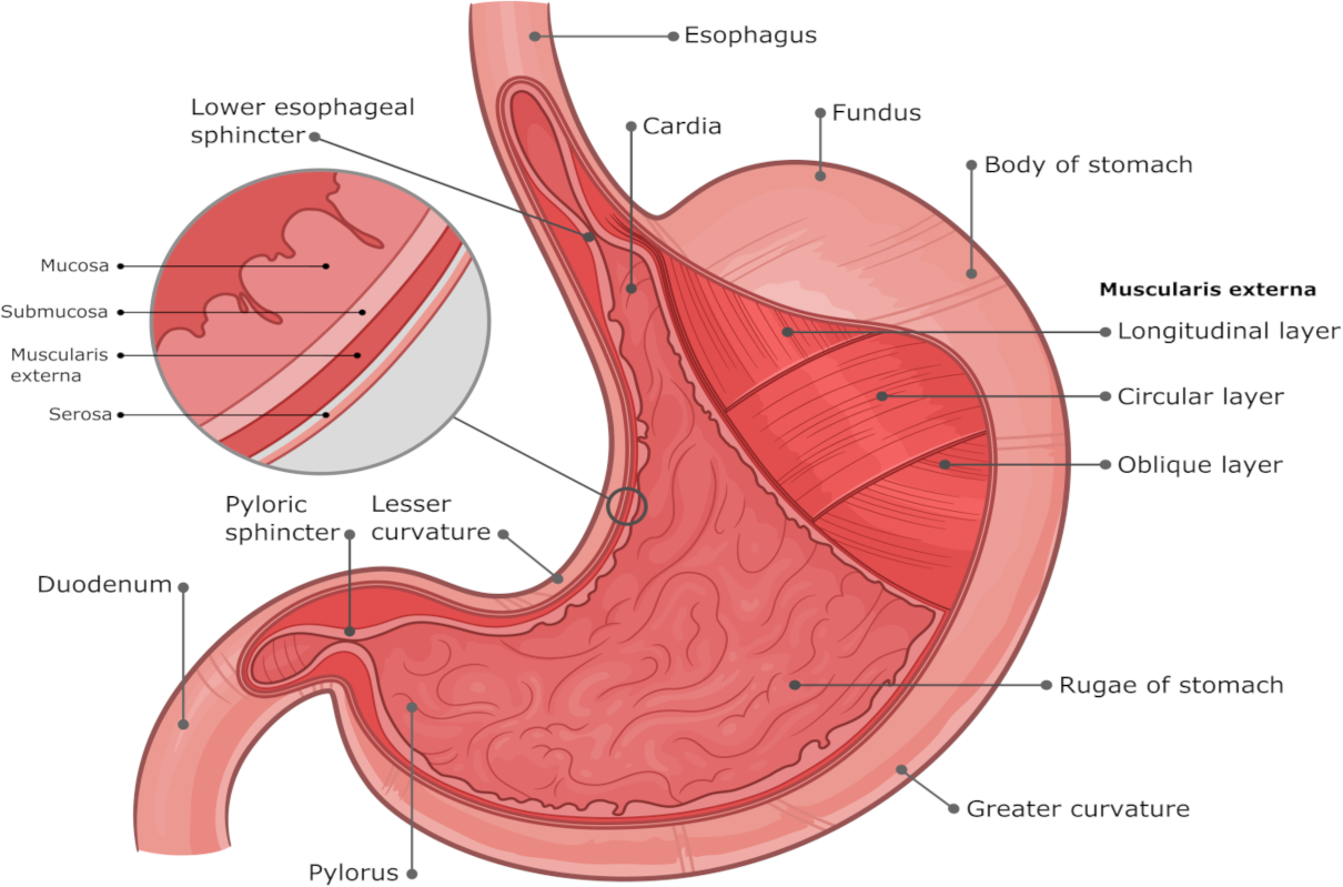
When looking at stomach anatomy, the organ is usually divided into four main regions: the cardia, fundus, body and pyloric region. Anatomists also describe the greater and lesser curvatures, the gastric wall, sphincters and the internal mucosal folds known as rugae.
The four regions of the stomach
The boundaries between the stomach’s regions are not always visible as sharp lines, but the four-part division is useful for describing its anatomy and function.
1. Cardia
The cardia is the small region surrounding the point where the oesophagus enters the stomach. It lies immediately below the gastro-oesophageal junction.
2. Fundus
The fundus is the rounded upper part of the stomach. It rises above the level of the gastro-oesophageal junction and lies beneath the diaphragm.
3. Body
The body forms the largest central part of the stomach. It is the main area in which food is stored temporarily and mixed with gastric juice.
4. Pyloric region
The pyloric region of the stomach forms its lower, narrowing portion. It includes the pyloric antrum and pyloric canal and ends at the pyloric sphincter, which controls the passage of stomach contents into the duodenum.
Greater and lesser curvature of the stomach
The characteristic J-shape of the stomach creates two curved borders.
Greater curvature
The greater curvature is the long, convex outer border of the stomach. It extends from the upper part of the stomach around its left and inferior aspect towards the pylorus.
Lesser curvature
The lesser curvature of the stomach is the shorter, concave border on its upper and right side. It runs between the cardia and pyloric region.
What does a stomach cross section show?
A stomach cross section shows that the organ is built from several specialised layers rather than being a simple hollow sac. From the lumen outwards, the principal layers are the mucosa, submucosa, muscularis externa and serosa.
Mucosa
The innermost lining contains gastric pits and glands that produce mucus, acid, enzymes and regulatory substances.
Submucosa
This supportive connective-tissue layer contains blood vessels, lymphatic vessels and nerves.
Muscularis externa
The stomach has three main smooth-muscle orientations: inner oblique, middle circular and outer longitudinal. Together they help churn and mix the stomach contents.
Serosa
The serosa is the smooth outer covering of the stomach and forms part of the visceral peritoneum.
Longitudinal section of the stomach
A longitudinal section of the stomach follows the organ along its longer axis. It can demonstrate the relationship between the cardia, fundus, body and pylorus, as well as the wall, internal folds and narrowing towards the pyloric canal.
When the stomach is relatively empty, its mucosa forms temporary folds called gastric rugae. These flatten as the stomach expands after food enters.
The sphincters of the stomach
Two muscular control regions are particularly important at the entrance to and exit from the stomach.
Lower oesophageal sphincter
The lower oesophageal sphincter is a physiological high-pressure zone at the junction between the oesophagus and stomach. It is sometimes referred to as the cardiac sphincter. Its function helps limit the backward movement of gastric contents into the oesophagus.
Pyloric sphincter
The pyloric sphincter is a thickened ring of circular smooth muscle at the junction between the stomach and duodenum. It regulates how quickly partially digested stomach contents enter the small intestine.
Gastric glands and stomach secretions
The gastric mucosa contains specialised cells that produce the substances needed for digestion and protection of the stomach lining.
| Cell type | Main secretion | Role |
|---|---|---|
| Mucous cells | Mucus and bicarbonate | Help protect the stomach lining from acid and digestive enzymes. |
| Parietal cells | Hydrochloric acid and intrinsic factor | Acid creates the low-pH environment needed for digestion. Intrinsic factor is required for normal vitamin B12 absorption later in the small intestine. |
| Chief cells | Pepsinogen and gastric lipase | Pepsinogen is converted into the protein-digesting enzyme pepsin. Gastric lipase contributes to fat digestion. |
| G cells | Gastrin | Helps regulate gastric acid secretion and stomach activity. |
Hydrochloric acid and pepsin
The stomach normally maintains a strongly acidic environment, although the exact pH varies with food intake and other physiological factors.
Parietal cells release hydrochloric acid (HCl). The acid helps unfold dietary proteins, supports the conversion of pepsinogen into active pepsin and contributes to defence against many microorganisms swallowed with food.
Pepsin begins the breakdown of dietary proteins into smaller peptide fragments. Unlike the old description of a fixed stomach pH of exactly 1.0, gastric acidity changes during digestion and typically becomes less acidic for a period after a meal.
How stomach digestion is regulated
Gastric activity is controlled by a combination of nervous and hormonal signals. Digestion is often described in three overlapping phases.
Cephalic phase
The sight, smell, taste or anticipation of food can stimulate gastric activity through nervous pathways before food reaches the stomach.
Gastric phase
Food entering and stretching the stomach stimulates local nerve reflexes and hormone release, including gastrin, which supports gastric secretion and motility.
Intestinal phase
As stomach contents enter the duodenum, signals from the small intestine help regulate further gastric secretion and slow gastric emptying when necessary.
From the stomach to the small intestine
Food does not pass directly through the stomach unchanged. Muscular contractions mix it with gastric secretions and gradually produce a semi-fluid mixture known as chyme.
The pyloric region then meters small amounts of chyme through the pyloric sphincter into the duodenum. This controlled process allows the small intestine to continue digestion and absorb nutrients.
Only limited absorption normally occurs through the stomach lining. The small intestine is the principal site of nutrient absorption.
Stomach anatomy: frequently asked questions
What are the four regions of the stomach?
The four main anatomical regions are the cardia, fundus, body and pyloric region. The pyloric region includes the pyloric antrum and pyloric canal before the stomach joins the duodenum.
What is the lesser curvature of the stomach?
The lesser curvature is the short, concave border on the upper and right side of the stomach. It runs from the area of the cardia towards the pylorus.
What is the greater curvature?
The greater curvature is the long, convex outer border of the stomach. It follows the left and lower contour of the organ.
What is the pyloric region of the stomach?
The pyloric region is the narrowing final section of the stomach. It leads towards the pyloric sphincter and the first part of the small intestine, the duodenum.
What are the sphincters of the stomach?
The important control regions are the lower oesophageal sphincter at the entrance to the stomach and the pyloric sphincter at its exit. The lower oesophageal sphincter is sometimes called the cardiac sphincter, although it is primarily a physiological rather than a clearly defined anatomical ring.
What does a stomach cross section show?
A cross section shows the lumen and the main layers of the stomach wall: mucosa, submucosa, muscularis externa and serosa. It may also show gastric folds, glands, blood vessels and the different orientations of smooth muscle.
What is a longitudinal section of the stomach?
A longitudinal section is a view cut along the longer axis of the stomach. It can demonstrate the relationship between its regions and the way the organ narrows from the body towards the pylorus.
Interested in digestive and bowel wellbeing?
The stomach and large bowel are different parts of the digestive system. Colon hydrotherapy works with the contents of the large bowel rather than the stomach. If you are exploring bowel wellbeing, Parkland Natural Health provides consultation-led colon hydrotherapy at our Holborn clinic in Central London.
